It’s a common, often overlooked, moment. You’re brushing your teeth, glance in the mirror, and suddenly notice something slightly off about one of your pupils. Perhaps it appears larger, unusually shaped, or responds differently to light. Initially, it might feel like an oddity too minor to mention. Many adults, particularly older individuals, might quietly dismiss these subtle shifts, hoping they’ll resolve on their own. Yet, the truth is, even minor alterations in pupil appearance or function can sometimes be significant indicators that your eyes or nervous system require attention. Recognizing these early signals empowers you to make proactive and informed decisions about your well-being.
Before delving into the specifics, it’s essential to understand a fundamental aspect of pupils that many people overlook. They are far more than just the dark circles at the center of your eyes. Functioning as intricate windows, pupils offer a direct glimpse into the complex workings of your nervous system. By the end of this comprehensive guide, you’ll gain a deeper appreciation for the surprising insights your pupils can reveal about your health.
Why Your Pupils Offer Vital Insights into Your Health
While most people associate pupil size changes primarily with varying light conditions, this is only a fraction of their story. The pupil’s dynamic adjustments are orchestrated by a sophisticated network of nerves that connect your eyes directly to your brain. When light enters the eye, signals are transmitted via the optic nerve and brainstem, triggering an automatic adjustment in pupil size. This involuntary reflex is crucial for protecting the delicate retina and ensuring optimal visual clarity.
However, because pupil function relies on the seamless interaction between your visual system and neurological pathways, any disruption along this intricate route can manifest as changes in how your pupils look or react. Extensive research in the fields of ophthalmology and neurology consistently demonstrates that variations in pupil characteristics can sometimes reflect:
- Underlying structural differences within the eye
- Alterations or damage to nerve pathways
- Residual effects from previous injuries or trauma
- Presence of congenital eye conditions from birth
- Reactions or side effects from certain medications
It’s crucial to emphasize, however, that not every unusual pupil indicates a serious underlying condition. Many individuals possess harmless, benign variations that have been present since birth. Nevertheless, understanding the different categories of pupillary abnormalities is vital, as it equips you to discern when a change warrants professional medical evaluation. This understanding leads us to explore the specific types of pupil anomalies.
9 Key Types of Pupillary Abnormalities to Recognize
Some differences in pupil appearance are immediately apparent, while others may only be detected through a thorough eye examination. Below are several commonly discussed types of pupillary abnormalities in eye health education:
Corectopia
In this condition, the pupil is positioned off-center, rather than perfectly in the middle of the iris. It can result from developmental variations or structural modifications within the eye itself.
Polycoria
Characterized by the presence of more than one distinct pupil opening in the same eye. True polycoria is exceptionally rare and typically congenital, meaning it’s present from birth.
Dyscoria
Instead of a perfectly round shape, the pupil appears irregular or distorted. This can sometimes develop following an eye injury, surgical intervention, or inflammation inside the eye.
Aniridia
A condition where part or all of the iris (the colored part of the eye that controls pupil size) is missing. Individuals with aniridia often experience heightened sensitivity to bright light due to the eye’s reduced ability to regulate light entry.
Coloboma
Often described as a ‘keyhole-shaped’ pupil, this occurs when a segment of the eye’s structure fails to develop completely before birth.
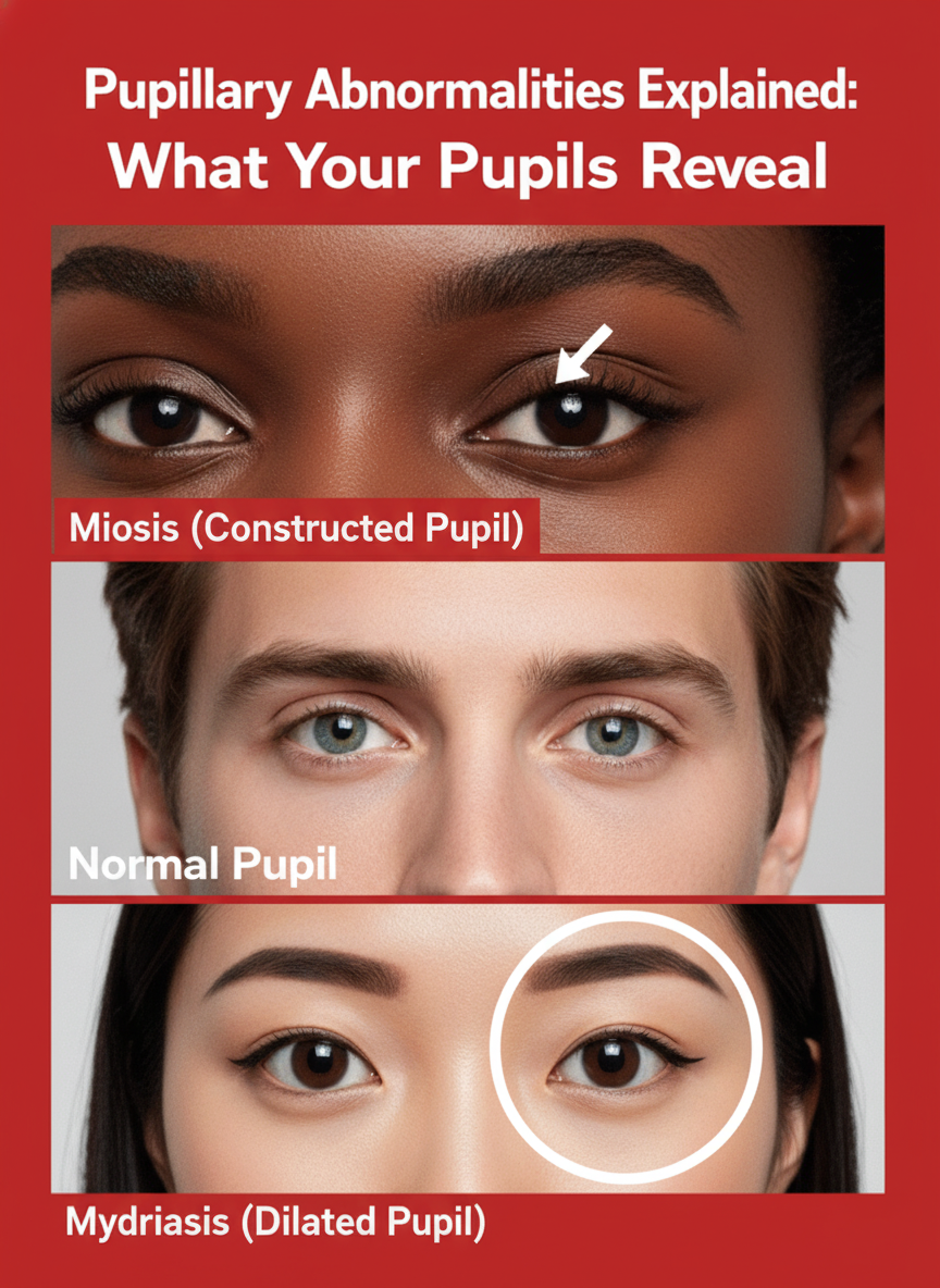
Permanent Mydriasis
The pupil remains abnormally dilated (large) even in brightly lit environments. This persistent enlargement can be caused by certain medications, specific neurological conditions, or eye injuries.
Permanent Miosis
Conversely, the pupil remains unusually constricted (small) and fails to adequately widen in dim lighting. This can sometimes be a normal part of the aging process or a side effect of particular medications.
Keyhole Pupil After Injury
Trauma to the eye or certain surgical procedures can physically alter the pupil’s shape, leading to a distinct ‘keyhole’ appearance. Eye care professionals closely monitor such changes to ensure normal eye pressure and function are maintained.
Marcus Gunn Pupil (Relative Afferent Pupillary Defect – RAPD)
This condition involves an abnormal pupillary response to light, typically signaling a problem with the optic nerve pathway. It’s often detected during a simple ‘swinging flashlight test’ performed by an ophthalmologist.
However, merely knowing the names of these conditions isn’t enough. What truly matters is recognizing the broader signals your body might be sending.
Accompanying Signs That May Appear with Pupil Changes
Sometimes, changes in the pupil are not isolated. Many individuals observe other subtle symptoms that emerge alongside them. Common accompanying signs that warrant attention may include:
- Unequal pupils (Anisocoria): One pupil is noticeably larger or smaller than the other.
- Sluggish reaction to light: Pupils respond slowly or inadequately when exposed to changes in light.
- Eyelid changes: Such as a drooping eyelid (ptosis) occurring on the same side as the affected pupil.
- Blurred or double vision: Any significant alteration in visual clarity.
- Persistent headaches: Especially those localized around the eyes or temples.
It’s an interesting fact that mild anisocoria, where pupils are slightly unequal in size, occurs naturally in approximately 20 percent of healthy individuals and is often completely harmless. However, any sudden, noticeable, or new onset of anisocoria, especially when accompanied by other symptoms, should never be dismissed. This leads us to the most critical section.
When Pupil Changes Necessitate Medical Evaluation
While many variations in pupil size or shape are benign and non-urgent, there are specific scenarios where prompt professional medical evaluation is crucial. Consider seeking immediate medical advice if you notice:
- Sudden onset of pupil difference: If one pupil abruptly changes in size or shape.
- Accompanying pain: Especially eye pain, headache, or neck pain.
- Vision changes: Such as sudden blurred vision, double vision, or vision loss.
- Drooping eyelid (ptosis): If the eyelid on the same side as the affected pupil suddenly sags.
- Recent head or eye injury: Any pupil changes following trauma should be checked.
- Light sensitivity or difficulty adapting to light: New or worsening symptoms.
- Signs of neurological distress: Such as dizziness, weakness, or difficulty speaking.
Your pupils are truly remarkable structures, offering a unique window into your overall health. By paying attention to these subtle signals and understanding their potential implications, you can proactively safeguard your vision and neurological well-being. When in doubt, always consult with an eye care professional or physician for an accurate diagnosis and appropriate guidance.